
In conversation with Peter Tepe | Section: Interviews
Abstract: In conversation with Peter Tepe, Oliver Thie speaks in detail about his project Oliarus polyphemus. Part I discusses: previous science-related works – his cooperation with the Natural History Museum, Berlin – working with the scanning electron microscope – a collage showing the entire animal in thousandfold magnification for the first time – experts’ reactions.
Part II deals with: our understanding of “the draughtsman’s interpretations” – an interest in things unnoticed by science – entomologists’ perspectives on the drawings – the self-identification as a researching draughtsman – the relationship to artistic research.
The draughtsman’s interpretations
Am I right in saying that as a draughtsman, you capture by hand what your selection of SEM recordings show?
An SEM recording in and of itself is the measurement result of a machine, presented as a collection of pixels in different grey tones. What the recording actually shows us can only emerge through observation and interpretation.
You also mentioned interpretations, or readings, in a panel discussion that took place on the last day of the exhibition in Düsseldorf. I would like to clarify what you meant by that. First and foremost, an SEM recording is “the measurement result of a machine”. Do you — like the entomologists — assume that the measurement result should be perceived as information about the structure of a part of the cicada?
To fundamentally consider the measurement result as information about the structure of parts of the cicada forms the premise for accepting the recordings as legitimate sources, enabling us to work with them. However, the mere presence of a recording does not translate as a statement about the cicada’s structure, because the machine takes measurements without knowing what it is measuring. This is where interpretation becomes a decisive factor.
What does this interpretation entail?
First and foremost, it means developing an assumption based on what the recording shows through close observation. Of course, entomologists do this too, however their perspective is shaped by scientific expectations and conventions. I operate in the belief that I am able to take a more unbiased approach here, or at least explore more aspects.
In addition to this, I enter a drawing dialogue that hugely intensifies my observations of the recording: drawing serves as an instrument to examine the visible, which sharpens my perception. Through the interplay of looking, depicting and comparing, I become conscious of details that remain invisible on a superficial level of observation. Only then can an interpretation truly take shape.
Finally, since both recording and drawing are visual complexes, drawing allows the developed conjecture to be articulated more precisely and directly than would be possible through language.
You make an important point here — to clarify it further, I’d like you to use a before-and-after comparison to demonstrate how you may have interpreted a concrete “measurement result of a machine, presented as a collection of pixels in different grey tones” by means of drawing.
I started by dividing the giant microscope-collage with a fine grid into squares measured 20×20 cm. Like the coordinates on a geographical map, I could now name each place on the image. With this orientation guide, I embarked on my drawing expeditions. Based on ever-changing research questions, I devoted myself to individual features and dealt only with those areas of the collage that were relevant to them. So it happened that I examined the same areas multiple times, but interpreted them quite differently. I’ll show you an example.
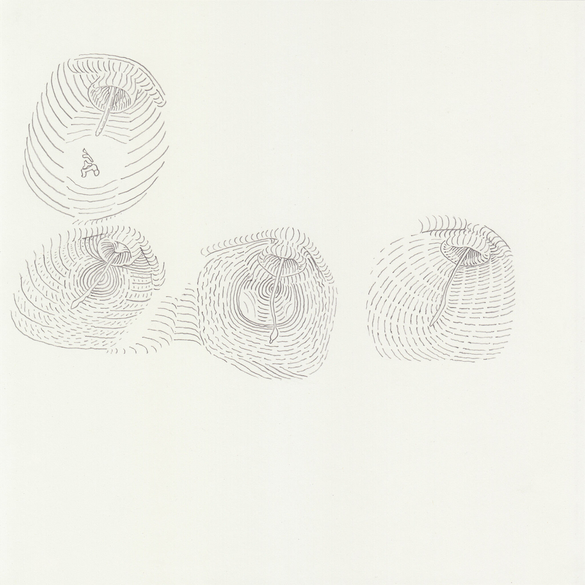
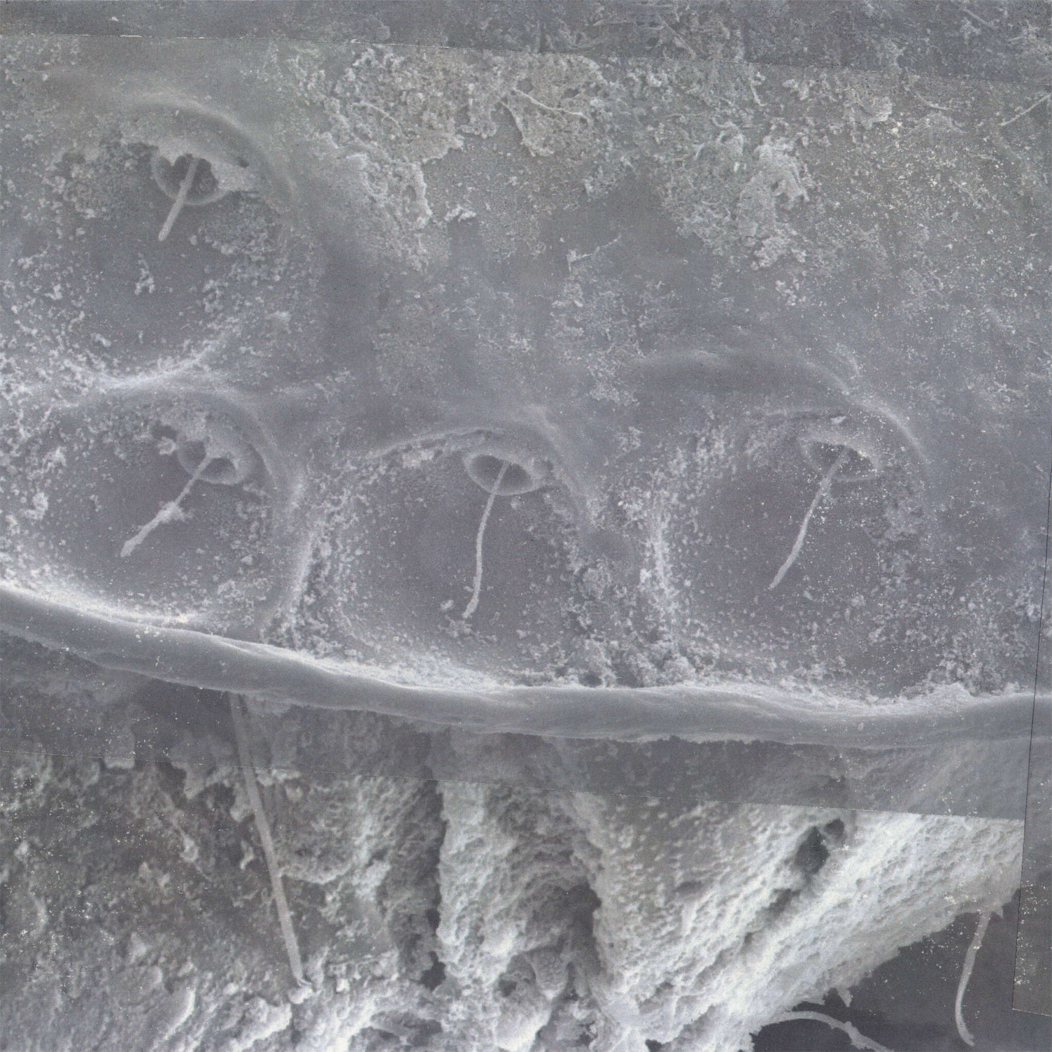
The upper side of the cicada bears a multitude of small pits, each of which has a single hair draped over it. The scientists assume that these form a sensory organ. However, its functionality is still unclear, which is also down to the fact that so far, it has not been possible to examine the overall formation via the microscope’s cropped field of vision. Expedition No. 3 frees the object of study from its rich visual surroundings and enables a clearer view. The drawing also defines the dimensions of each rather contourless pit. I chose to use curved lines as a graphic tool that allows the contemplation of volume.
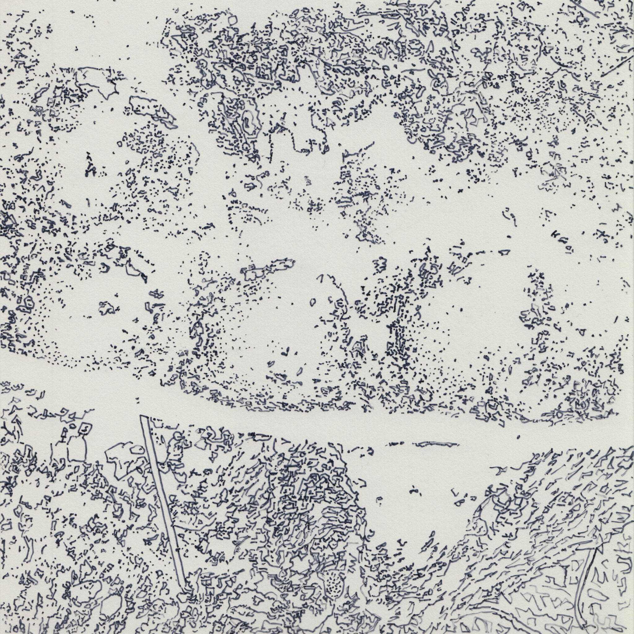
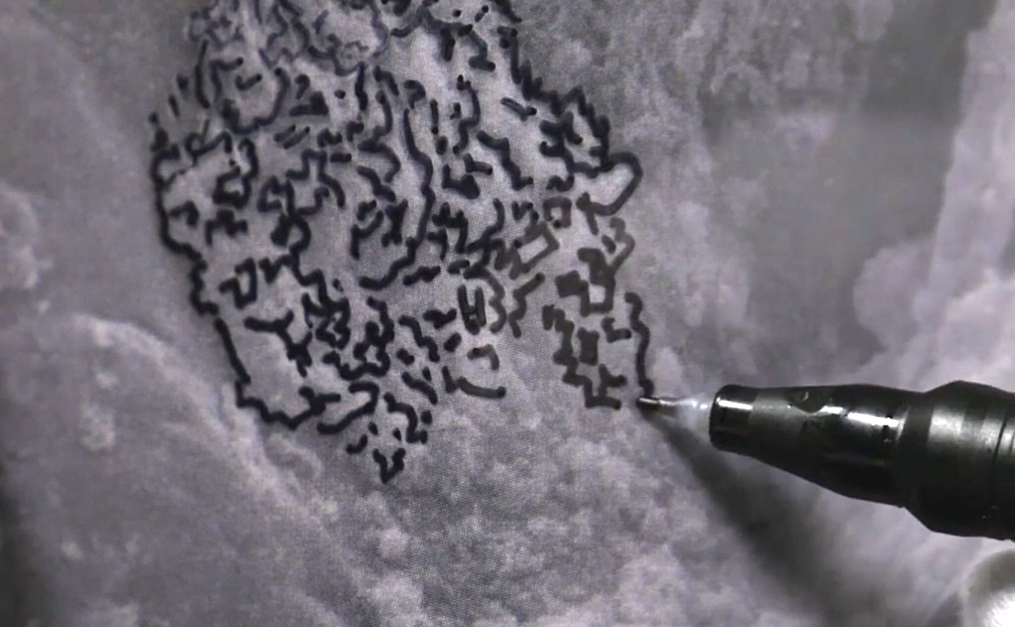
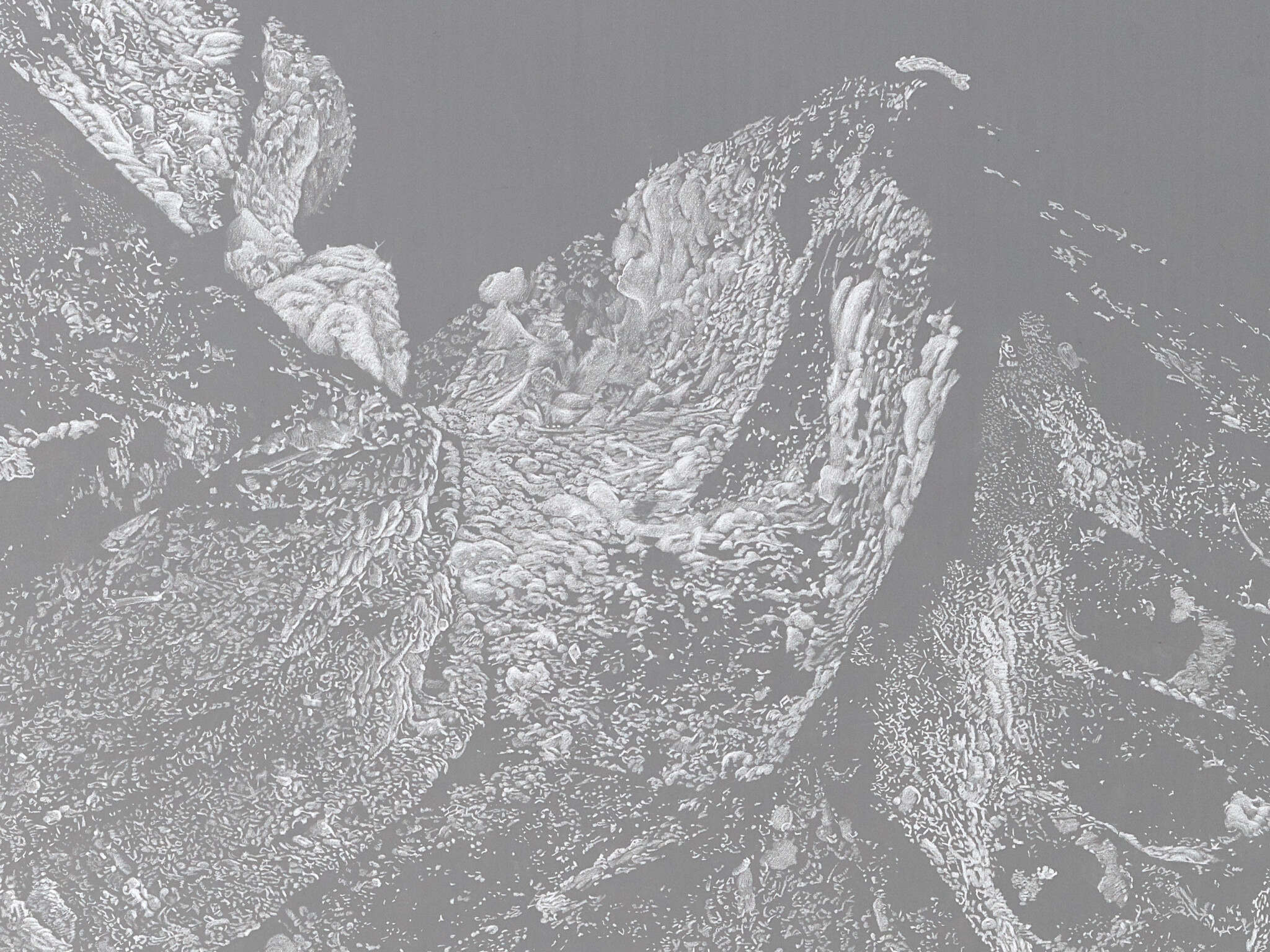
The surface of the cicada is covered in crumbs, fluff and dust. In conversation with the entomologists, I couldn’t help but notice that they completely disregard these ubiquitous deposits — assuming they were just dirt. My interest in things overlooked by science led me to make Expedition No. 6: I tried outlining everything supposedly external along a longitudinal section. Millimetre by millimetre, I traced the recording onto a directly applied film. In doing so, I became aware of the ambiguity of the technical image. Trying to capture what was shown here through lines required many guesses. Even though I was tracing the image, it is impossible to produce the same drawing twice: instead, it becomes a kind of subjective progress report that says “as I encountered this spot, it seemed to me to look like X or Y”.
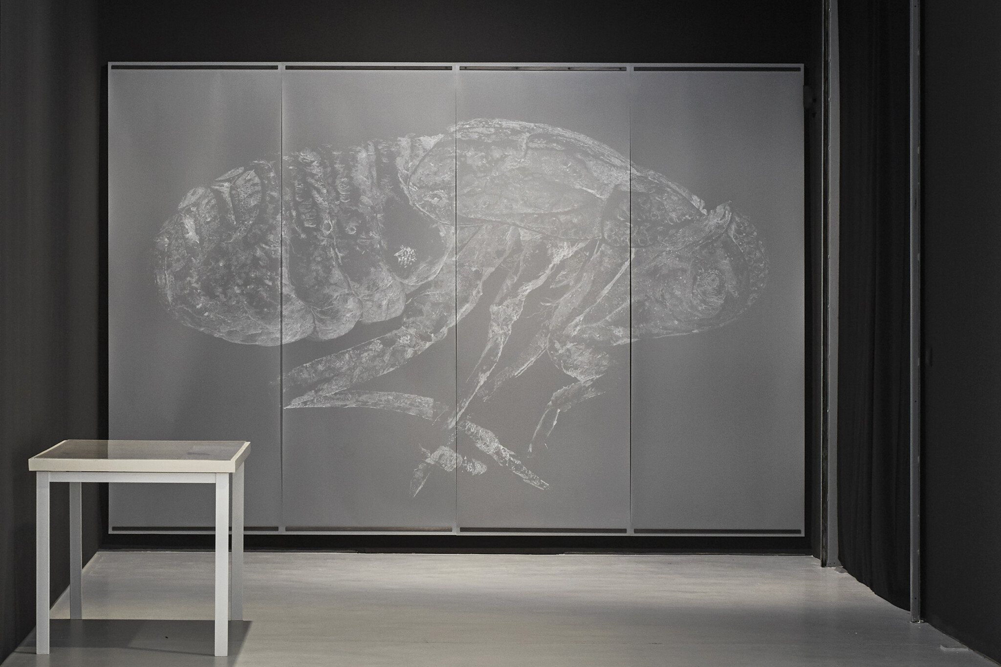
Incidentally, my interest in those crumbs on the cicada never left me — which led to the work that you saw in Gulliver’s Sketchbook. In Expedition No. 10, I explored for the first time the entire image. To get an even more accurate depiction of the crumbs and debris in this instance, I used a white pencil crayon as an overlay to somewhat remodel them. When the transparent drawing becomes visible against a black background, the deposits stand out from the body of the animal — although it remains clearly visible, the work actually depicts everything but the cicada itself.
So you understand “interpretation” to mean the following: faced with the visual fullness of an SEM recording, you concentrate on one specific characteristic and capture it through drawing; these drawings make a statement which is to be understood as a progress report with speculative elements. In other words: “interpretation” as you see it consists of reading an SEM recording, deciphering it — for example as a dent of some sort —and then translating it through drawing.
That’s right, although there are also investigations that go beyond the interpretations of SEM recordings, combining information from different sources. These create images that can be understood as models and depict something that no imaging technique alone can express.
An example would be great.
Of course.
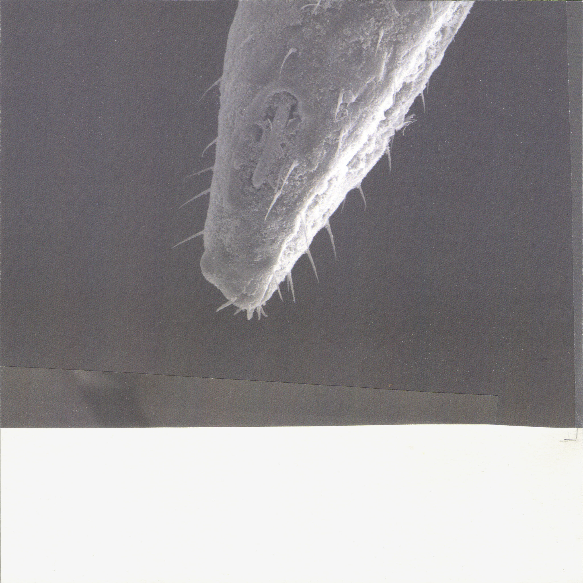
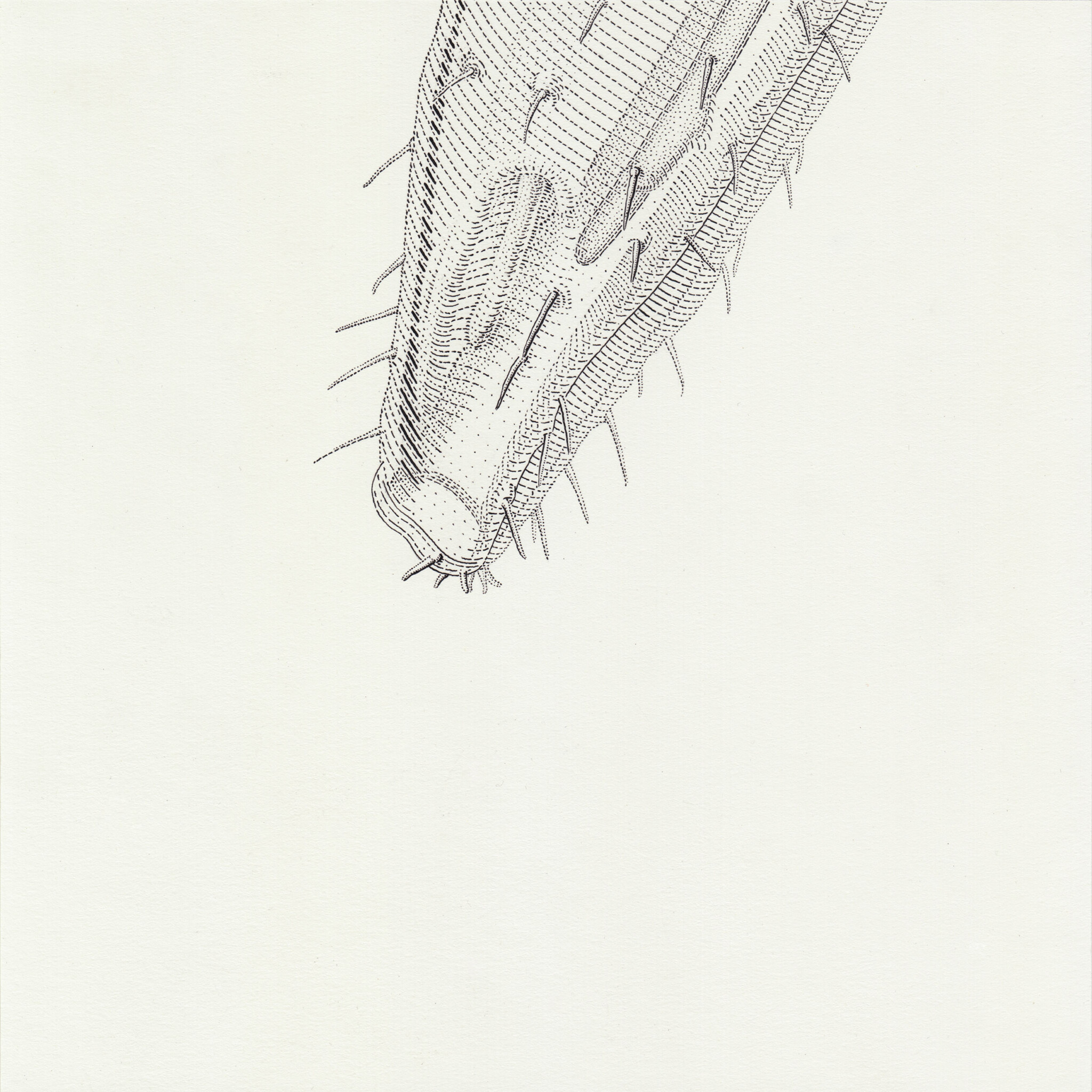
The electron microscope cannot reproduce colours, and transparent qualities also get lost. Everything appears opaque and impermeable. However, thanks to the optical microscope, I knew the cicada was almost translucent in many areas, especially its delicate limbs and proboscis. Inside the proboscis rests a spike, which the insect pushes out to suck plant sap. In Expedition No. 8, I illuminate the surface structures provided by the SEM and add a sense of transparency to them.
What do the entomologists with whom you are collaborating think about your drawings? Do they consider them insightful for the entomological cognitive process?
My investigations involve various forms of exchange with the scientists. Sometimes I take up something in their interest, other times I go in the opposite direction — so their reactions vary accordingly. Expedition No. 3 was met with a concrete response, because a certain feature that had only ever been observed in parts could be viewed as a whole for the first time, which also revealed the systemics of its configuration. Moreover, my drawing translation enabled the entomologists to gain insights on the individuality of each pit, which led them to reflect on their tendency to schematise impressions perhaps too quickly.
Expedition No. 6 prompted the entomologists to arrive at the general resolution not to subtract too much from what is actually visible. This brought us to the more philosophical question of where an organism ends and the surrounding environment begins, and realised that this boundary becomes increasingly blurred in the microcosm.
Now that we have discussed your understanding of “interpretation”, let’s talk about your self-identity as a researching draughtsman. A conventional scientific illustrator produces, for example, drawings of insects newly discovered by scientists. These drawings must meet a certain criteria that are determined by the scientific research process. How does a researching draughtsman stand in contrast to this? Do they participate in the scientific cognitive process or is their activity defined according to other criteria?
Barbara Wittmann’s text Zeichnen am Mikroskop (Drawing at the Microscope) nicely points out that drawing can be an essential part of the scientific research process: it advances morphological conceptualisation by enabling us to recover and articulate forms of organisms from the unknown. My work, too, grounds itself in the self-conception of this epistemic craft — the main difference being that I can pursue broader interests within my work without having a scientific mandate to obtain certain results. I initiate an independent epistemological process that runs parallel to the scientific process, wherein I examine the respective subject matter in a different way.
So, currently you are not interested in making the generic distinction between the researching draughtsman and the scientist — rather, you are concerned with differentiating between two different ways of studying the cave cicada: both of which rely on the Scanning Electron Microscope and aim to expand knowledge on the animal’s composition:
Form 1: The entomologists follow the procedure that is typical for this science, as mentioned above.
Form 2: The researching draughtsman pursues the same goal but by other means: during the drawing process, they develop hypotheses based on the SEM results about the composition of parts of the cicada.
Rather than simply partaking in the scientific cognitive process, the researching draughtsman initiates an independent epistemological process. We are thus dealing with two different styles of research.
Yes, as you say, I would like to increase our knowledge on the nature of the cave cicada. Overall, however, I am interested in expanding scientific access to the world. Scientific topics and procedures open up spheres of research to me that I would not be able to reach otherwise, which then also become subject of my own research. By studying them in detail — identifying previously unnoticed aspects and developing my own methods to address them — I also thematise the scientific approaches themselves. In order to keep this process going, I constantly immerse myself in different contexts and collaborate with different disciplines.
Last question: as you consider yourself a researching draughtsman, it seems natural to assume that you also identify with the diverse field of artistic research — a topic which is discussed in various w/k contributions. If this is the case, please share your own understanding of this guiding principle.
As I understand it, artistic research is characterised by using artistic means to gain insights into a subject matter that lies outside the traditional domains of art. My position has gradually and naturally developed in this direction. When I consciously started drawing in order to gain a deeper understanding of phenomena in the outside world and to investigate the mechanisms of my own perception, I discovered such an artistic research tool for myself. Then there was my close relationship to the natural sciences. I’m not sure if artistic research always needs to be defined in direct comparison to science. However, since scientific questions and methods became my source of inspiration, I have oriented myself towards the aforementioned attributes of a research-oriented approach. So I too follow a methodology that was developed experimentally and then established, which, when applied repeatedly, leads to serial works. Moreover, I strive for forms of publication that will communicate the insights gained, disclose contexts and methods, and make them available for further discussion. Despite my affiliation to the (natural) sciences, there are also many differences. I do not aim for quantitative results or draw general conclusions. I am much more interested in the individual qualities of each case and in the experience of the research process. With the drawings I keep all my experiments visible: observation, perception and wonder remain present.
Oliver Thie, thank you for this insightful interview. A follow-up is planned in which we will explore your unique position in the interplay between science and art in more detail, also taking into account the most important stages of your artistic development.
Literature
Thie, Oliver (2016): Oliarus polyphemus. Documentary. Online: https://vimeo.com/165158411
Wittmann, Barbara (2013): Morphologische Erkundungen. Zeichnen am Mikroskop. In: Bildwelten des Wissens 9.2: Morphologie, pp. 45–54.
Details of the cover photo: videostill from the documentary: Oliver Thie — Oliarus polyphemus (2016).
Translated by Rebecca Grundmann.
How to cite this article
Oliver Thie and Peter Tepe (2023): Oliver Thie: Drawing as Research. Part II. w/k–Between Science & Art Journal. https://doi.org/10.55597/e9137

Be First to Comment