In conversation with Peter Tepe | Section: Interviews
Abstract: In conversation with Peter Tepe, Oliver Thie speaks in detail about his project Oliarus polyphemus. Part I discusses: previous science-related works – his cooperation with the Natural History Museum, Berlin – working with the scanning electron microscope – a collage showing the entire animal in thousandfold magnification for the first time – experts’ reactions.
Part II deals with: our understanding of “the draughtsman’s interpretations” – an interest in things unnoticed by science – entomologists’ perspectives on the drawings – the self-identification as a researching draughtsman – the relationship to artistic research.
Oliver Thie, in this interview we will primarily focus on your current project Oliarus polyphemus – Drawing Expeditions in the Microcosm. Parts of this project were shown in the exhibition Gulliver’s Sketchbook at the Arthena Foundation in Düsseldorf from 10 March – 26 June 2022.
w/k is also interested in previous science-related works. To begin with, you can give us a brief summary of these projects. This will also help us contextualise Oliarus polyphemus in your artistic development thus far.
Before and after beginning my work on Oliarus polyphemus, I accompanied several biological expeditions where I recorded my own observations, for example with Buteo buteo (2013) on the flight movements of the Eurasian Buzzard in Nordeney or with The Social Aphid Experiment (2016) on behavioural patterns of social aphids in the US. In 2018, together with historian Angela Strauß and media scientist Sebastian Döring, I established Das Temporäre Objektlabor (The Temporary Object Laboratory): a workshop for researching scientific collections, located publicly in the Tieranatomisches Theater (Veterinary Anatomy Theater), a museum in Berlin. In this context I developed the Schattenfänger-Methode (shadow-capturing-method). Since then, I have been exploring enlarged silhouettes of natural objects with soot drawings. By 2020 I had created Die Wahrheit über den Ursprung der Welt (The Truth about the Origin of the World); shadow images of a historical rock collection from the Siebengebirge, which is currently stored in the Natural History Museum, Berlin.
That’s a great deal of artistic projects relevant to w/k that we could explore in future articles. Now to our main focus: what is Oliarus polyphemus about? We also want to address those who are interested in the major topic of art and science or in certain aspects of it, but who have no prior knowledge of biology.
Cooperation with entomology
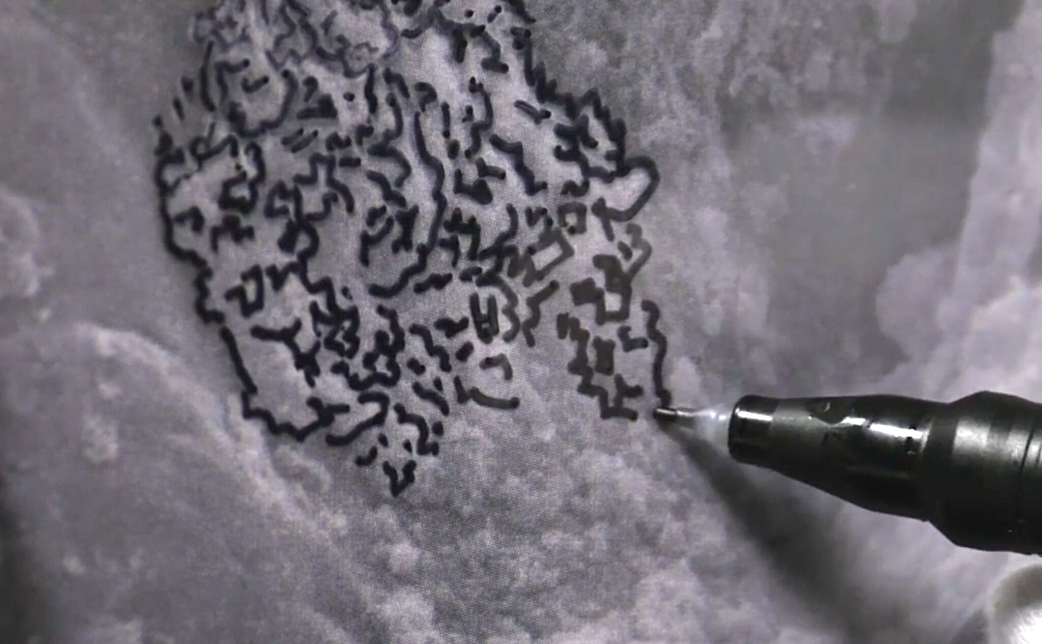
My artistic work often revolves around the expansion of perceptions through magnification. Oliarus polyphemus is the scientific name of the Hawaiian cave-dwelling cicada, an insect only three millimetres wide. Since 2014 I have been observing a specimen through the electron microscope magnified one thousand times, exploring its surface structure by means of drawing.
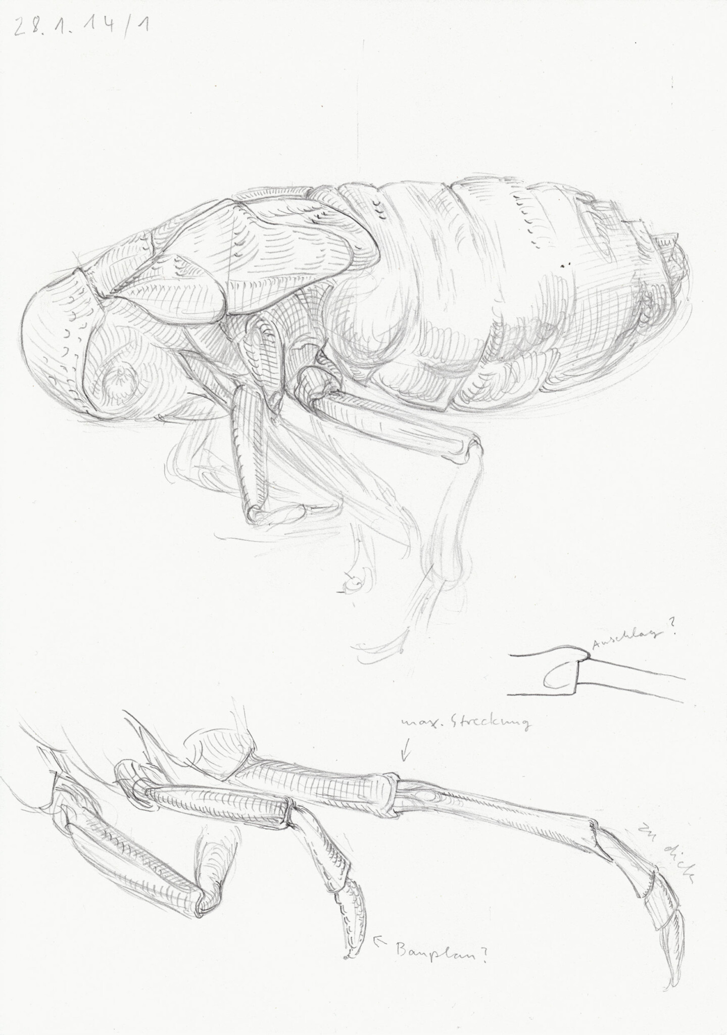
In what setting did – and does – this work take place?
Based on the proposition of magnifying an insect, I was able to enter a cooperation with the entomologists Prof. Dr. Hannelore Hoch and Dr. Andreas Wessel from the Natural History Museum, Berlin. They suggested the cave-dwelling cicada because these animals exhibit diverse characteristics that have not yet been studied fully and are object of their ongoing evolutionary research. Besides, the cicadas are small enough to subject them to a thousandfold magnification, which means that three millimetres become three metres. Using larger animals would have quickly overwhelmed the project. From 2014 to 2016 I obtained the status of a guest scientist, which enabled me to work in the Natural History Museum. During this time, I ran a public drawing lab in the exhibition area. After completing the residency, the project was temporarily put to rest. In 2021 I took it up again and am now continuing to work on it in my studio.
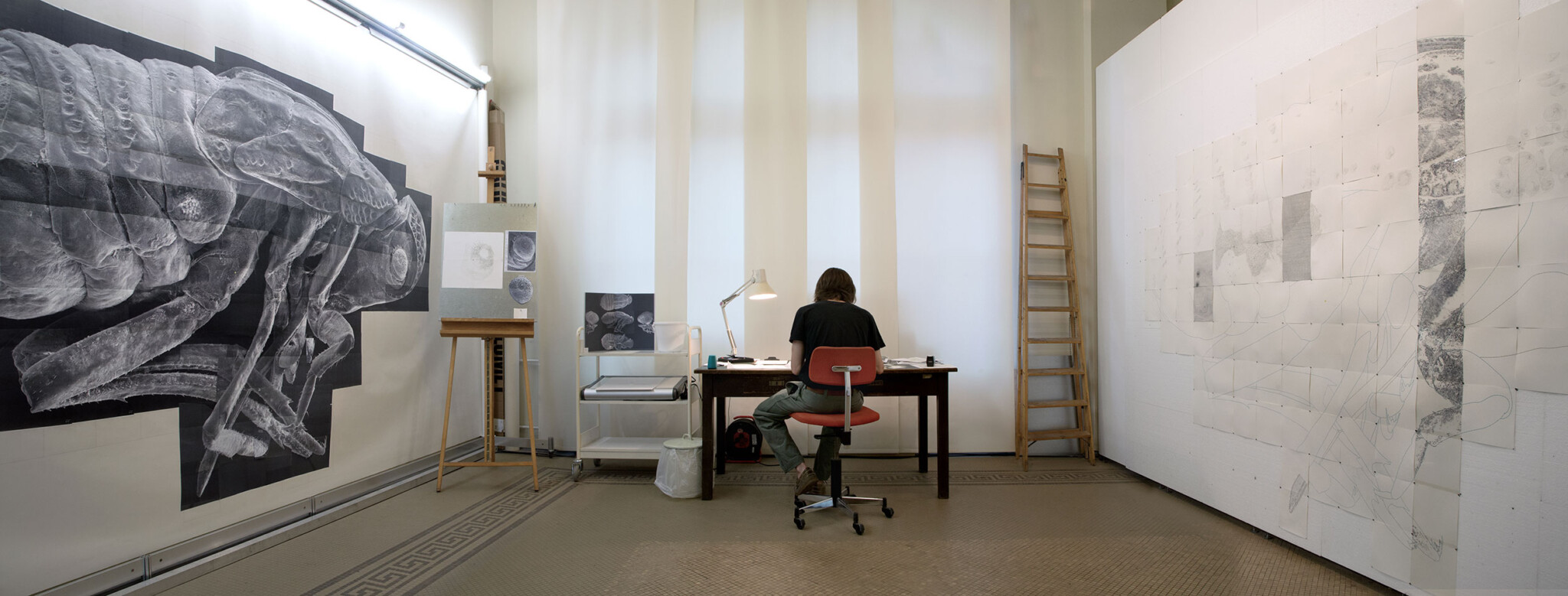
How did the cooperation with entomology come about?
Before I started doing independent research as an artist, I worked as a scientific illustrator. For my future collaboration partners, I made illustrations of newly discovered insects that were then published together with their species description in scientific journals. As time went on, however, I developed research interests that I was not able to explore within a contractual work relationship. Equipped with my own funding and on the basis of the mutual trust that had developed over the years, I was able to gradually transform my role as supplier into that of a collaboration partner.
What did you do when you first started the project?
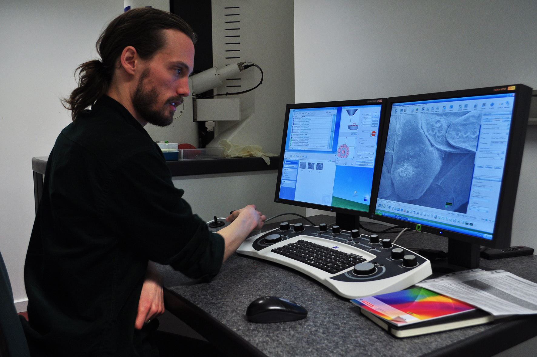
To obtain a thousandfold magnification, you have to work with the scanning electron microscope (SEM). At the Natural History Museum I had access to this technology and was able to learn how to use it. It’s not like a conventional microscope, where you lay something beneath the lens and look through it. The sample has to undergo an elaborate preparation in order for it to even be viewable in the first place. It is then placed in an insulated vacuum chamber and scanned with a beam of electrons, whose interactions with the sample are transformed into an image by the computer. Next to it is a monitor which shows the result. A joystick is used to move the sample and a rotary knob is for zooming in. However, these functions reveal a fundamental problem of magnifying instruments: the monitor can only show a small part! Due to the limited screen size, the overview of the body is lost and the possibilities of gaining knowledge in the microcosm remain fragmentary.
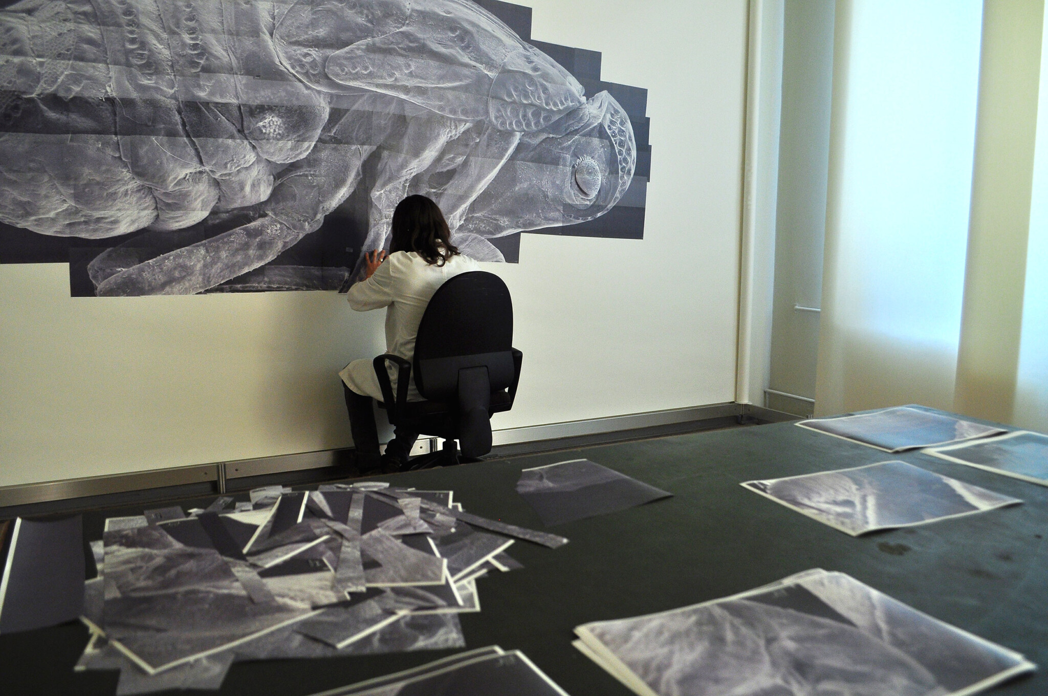
With the aim to overcome this, I systematically went over the entire body of the cicada through the SEM, capturing hundreds of pictures to document each section. These I made into a collage, making it possible to view the whole animal in thousandfold magnification for the first time. But because the collage is based on computer generated images that do not yet contain a semantic proposition, it did not yield a research result. Even as a scientific illustrator I worked on articulating visual analyses through drawing. Now I wanted to explore this potential further, to gain actual insights from the microscopic image.
One of my goals is to adequately situate what you do in Oliarus polyphemus with help of the terms and theoretical assumptions used in w/k. In particular, I am interested in explicating your concept of research-drawing and its background beliefs in simple terms. The first two points are indisputable:
(1) You, like several entomologists, are studying the three-millimetre Hawaiian cave cicada.
(2) With the help of the scanning electron microscope, you, like many scientists, obtain thousandfold magnifications of certain features of the cicada, such as a particular pit.
Now to classify these magnifications:
(3) The SEM allows for our descriptive knowledge to expand: the cicada’s physical structure is captured more precisely than before. This is to be distinguished from the expansion of knowledge by theory-based explanations.
Yes, that is correct.
(4) Whilst some entomologists pursue their work in a specialised manner and remain content with closely examining specific characteristics of the cicada revealed by the SEM, you are interested in capturing the cicada as a whole. To achieve this, you used “hundreds of SEM-recordings” to create a collage, “making it possible to view the whole animal in thousandfold magnification”. Does your interest in an overall view of the cicada have anything to do with the fact that you are an artist? Or, in other words, can the making of a collage be understood as an artistic intervention?
That’s how I’d see it. My primary aim was to make it possible to view the animal in its entirety, which also meant that many of the images I captured would not have been of scientific interest. On the one hand, this is because as single, isolated images they may be unclear and only reveal their meaning as part of the puzzle. On the other hand, because many areas are not considered to have details of scientific relevance and are therefore skipped over. In contrast to this, I proceeded without any particular expectations and simply captured what I could find. Also, the analogue approach of assembling images with scissors and glue is untypical in the technologically advanced scientific world – it much rather ties in with a pictorial tradition.
I would like to expand on point (4). The desire to capture the phenomenon in question as a whole also exists in the empirical sciences – so surely not all entomologists take an exclusively specialist approach. How was your collage received in the team? Have there been other attempts to capture the animal in its entirety that stem primarily from a scientific context?
Initiatives to digitise entomological collections have been on the rise for some years now. The Natural History Museum in Berlin, for example, is running a pilot project to test special 3D scanners used to depict individual animals. However, these scanners lack in resolution and thus do not achieve SEM-like magnifications. It is quite common in scientific procedures to study the animal as a whole, albeit within a lower magnification range. In the higher ranges, it quickly becomes too technically complex.
The problem of the cropped field of vision was something my collaboration partners were well aware of, but alongside their daily work there was simply no room or necessity to work on solutions. Dr. Wessel is the only biologist I know of who, before our collaboration, had already made an attempt in this direction by digitally assembling a structure from several individual images. However, the huge volume of image data, and the fact that an image created this way must first be printed out in order to be viewed without restriction, set technical limits to this process. My collage method was the only viable way for me to assemble the masses of data, allowing for a coherent whole to emerge. A physical relationship to the animal was created, which was a completely new experience for the entomologists – its structures could be perceived throughout its entire body, without having to use optical instruments. You feel like you’ve been transported to the microcosm.
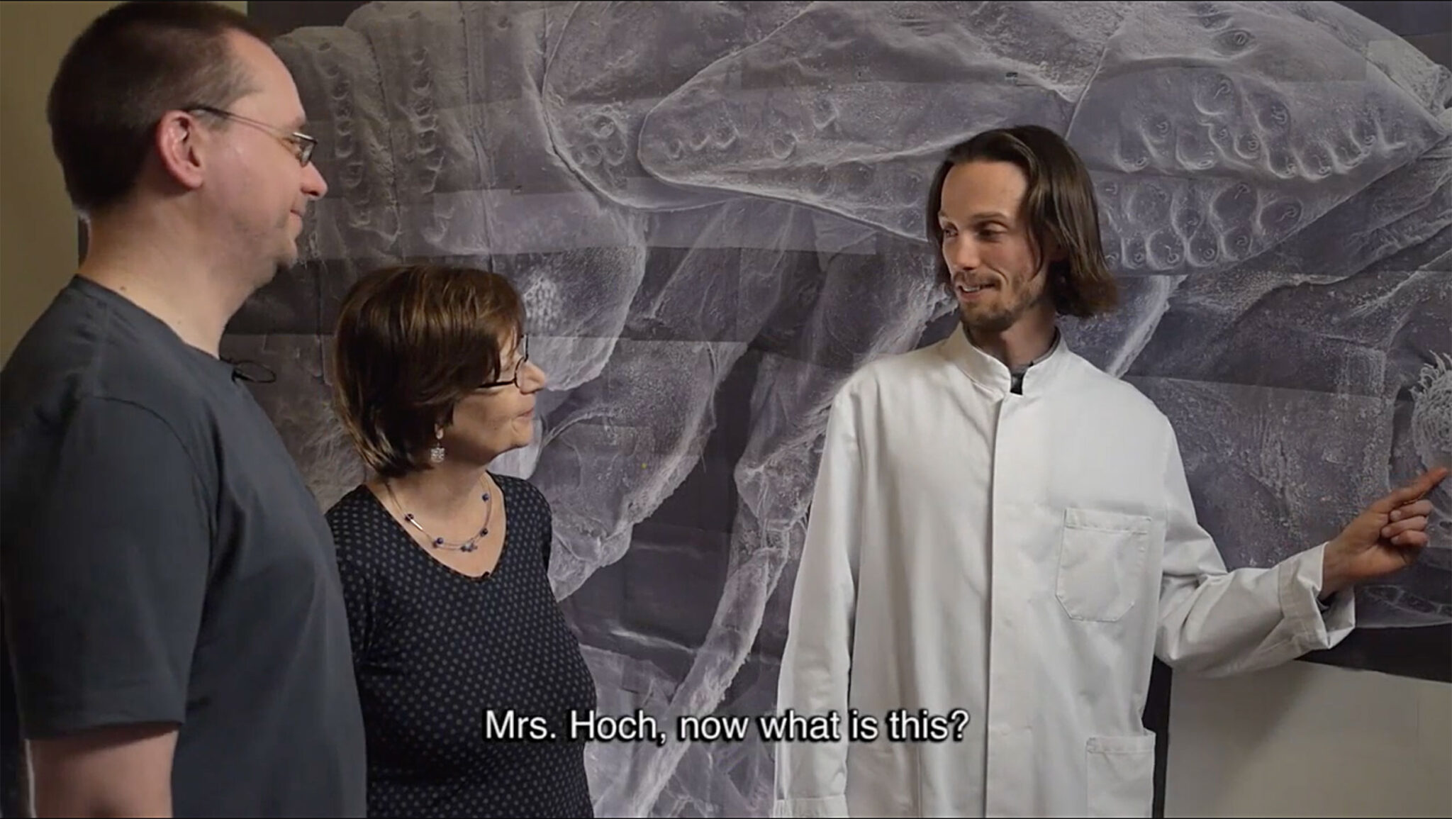
To summarise: there is also interest amongst entomologists in an overall depiction of the cicada’s microstructures, but for technical reasons this interest is yet to be fulfilled.
Literature
Thie, Oliver (2016): Oliarus polyphemus. Documentary. Online: https://vimeo.com/165158411
▷ Continue to part II of the interview
Details of the cover photo: videostill from the documentary: Oliver Thie – Oliarus polyphemus (2016).
Translated by Rebecca Grundmann.
How to cite this article
Oliver Thie and Peter Tepe (2023): Oliver Thie: Drawing as Research. Part I. w/k–Between Science & Art Journal. https://doi.org/10.55597/e9121

Be First to Comment