

Text: Hubert Mayer | Section: Art-related Science
Summary: On the occasion of the exhibition Cell Vitality, Hubert Mayer first gives an introduction to genetic engineering and then discusses a project of art-related science: three researchers, with the help of artistic media, explain the findings of their discipline in order to convey these to a wider general public.
The exhibition Cell Vitality took place in the Braunschweig Botanical Gardens. Before discussing the exhibition concept with Peter Tepe, I will begin with a short and simple introduction to genetic engineering and then briefly address the topics science communication and science-related art.
Genetic Engineering as a Science
The term genetic engineering can be considered synonymous with biotechnology, molecular genetics and molecular biology. It has developed exponentially in a short time from an isolated discipline into an interdisciplinary field of science. It is a procedure that aims to specifically intervene in the genetic material (gene) and thus in the biochemical regulation (proteins) of cells in living organisms. Targeted gene modification is already being tested in animal and plant breeding. Initially, it was used for scientific and technical purposes in biotechnology for the production of biological substances, but more recently it has also played an important role in health research, such as the production of vaccines against the SARS-CoV-2 virus.
Genetic engineering is based on a knowledge of cell biology and makes use of important cellular building blocks, i.e. biopolymers. Biopolymers are long chains of monomeric units that build complex structures and can carry out various functions. The most common example of this is our hereditary substance, the DNA (deoxyribonucleic acid), which is the general carrier of a cell’s genetic information. The DNA consists of a double strand of the complementary nucleobases A – T and G – C.
The mRNA (messenger ribonucleic acid) is the mobile carrier of information. It mainly carries the information of just one singular gene and thus only a tiny part of a cell’s total genetic information. In contrast to DNA, mRNA is single-stranded, but still consists of nucleobases. The base sequence of an mRNA can be translated into a protein. Proteins consist of chains of amino acids and, if folded correctly (into a 3D structure), can carry out various functions; they are the cell’s so called workhorse. The goal of genetic engineering is to reinterpret the central dogma of molecular biology, that is, the natural direction from DNA to mRNA to protein, and to define and use the flow of information forward and, to some extent, backward.
The model of DNA as a double helix presented by J. Watson and F. Crick in 1953 is considered the central starting point. The process of deciphering the total sequence of the human genome led to a competition between two groups, which ended in the year 2000 with a compromise whereby both parties shared their results simultaneously. The entire human genome consists of 3.27 x 10⁹ building blocks (base pairs) with a total length of approx. 1.8 metres, all condensed together in 46 chromosomes within each individual cell. The exact number of genes, i.e. the units that code for mRNA and the proteins derived from it, is unknown. It has been established that there are between 80,000 and 140,000 genes. The genetic sequences of the most commonly inherited genetic illnesses are known, as well as those of most pets and useful plants. The DNA sequences of a large number of viruses, bacteria and fungi are also known. The information is used to produce genetic family trees that show the degree of kinship that can be used to identify the exact causes in the case of similar illnesses, e.g. SARS-CoV-2 and influenza viruses.
The central dogma for all cellular life (bacteria to mammals) allows simple organisms, e.g. bacteria, to be manipulated to produce human proteins. After isolating the corresponding human gene and transferring it into a bacterium, it is possible to produce unlimited amounts of the desired human product for medical use (eg. human insulin or blood factors). Bacteria can be manipulated to degrade environmentally dangerous and poisonous substances in a multi-stage biological process. The incorporation of plant-based gene-cascades enables the bacterium, like plants, to fix CO2 from the air and to produce carbon compounds: they are referred to as hybrids. These examples show that the genetic code is universal.
A great step forward in genetic engineering was achieved using a method for the duplication of DNA segments, the polymerase-chain-reaction (PCR). This procedure is indispensable, for instance, in forensics, genealogy, cultivation and virus identification (currently for the corona virus SARS-CoV-2).
The condition of a cell can be comprehensively characterised by the construction of gene expression profiles that record and characterise the entire mRNA profile at a specific point in time. The hope is that in complex diseases such as cancer, infectious diseases or neurodegenerative diseases, e.g. brain damage, it will be possible to recognise when a cell not only deviates from its healthy state, but also when it returns to it. Such analyses involving large data sets are only possible through the use of high-performance computers. Thus, genetic engineering can be thought of as a cross between the bio and computer sciences.
For the understanding of life processes, a targeted intervention in a cell, in an organism, is considered essential. The effect of a gene can only be studied in its respective cellular environment. As of late, the genetic scissors termed the Crispr/Cas 9 can be used to precisely modify the genetic material inside the living cell (Nobel Prize 2020). This system occurs naturally in bacteria, enabling them to cut up viral invaders. It has been modified to also function in higher organisms. A special feature of this is that any DNA section in a genome can be targeted by means of an appropriately designed guide (RNA). The nuclease, that is the enzyme that cleaves nucleic acids, is attached through a piggyback process to the RNA and then cuts the DNA at a specific position. The cellular repair system then closes the gap. In living cells, it can function to remove and/or replace particular units of the gene, which can then be checked by PCR and DNA sequencing. This technique is now widespread and is used in plant and animal breeding. The first clinical trials in humans who have a genetic defect in haemoglobin are underway (somatic gene therapy).
Embryonic development of an organism involves a highly complex dynamic: a fertilised egg cell, called an embryonic stem cell, develops into an organism. The total genetic information of the DNA remains the same in every body cell. Cell specificity occurs through activation of tissue-specific genes. Unravelling a corresponding DNA region is thus an important prerequisite. The tangled state (A) of each differentiated tissue cell should be different to that of an embryonic stem cell (B). This hypothesis has been tested in an animal model. The intact nucleus of a differentiated tissue cell (A) of a sheep was extracted, transferred as donor DNA into an enucleated egg cell (B) and delivered by a surrogate mother sheep. This resulted in the birth of a lamb: the clone Dolly. According to the above hypothesis, we can thus conclude that a transfer from A (differentiated) to B (undifferentiated) has taken place – similar to what occurs upon fertilisation of an egg cell. The process of asexual reproduction is known as cloning, i.e. identical reproduction. This is how bacteria usually reproduce. It is also widespread in plants, for instance in tubers, onions or cuttings of vine and fruit varieties. It is also still to be found in primitive vertebrates and some insect species.
Intact cellular functions are a characteristic of life, with proteins being the essential tools: Their three-dimensional structure contains highly specific functions, involving the creation and degradation of certain substances – the metabolism. Small changes in these structures can cause some functions to partially or completely stop working. Detailed structural knowledge is a prerequisite for the development of drugs, for example of antiviral substances. The drug’s active substance should fit exactly into the virus binding site (receptor) with high affinity and thus hinder the building of the virus. This type of drug can be administered in tablet form. Immunisation adopts a different approach, whereby a section of the viral envelope or a killed virus is inoculated into the body to activate the immune system, thus suppressing the invading process of the virus.
Science Communication
Genetic engineering has found application in many areas of modern society and created surprising innovations. Its use has elicited controversial views both nationally and internationally. During the fight against the SARS-CoV-2 pandemic, genetic engineering, with its biological knowledge, is the focus of international interest. Detection, spread and targeted reduction of the virus present an enormous problem worldwide. According to the WHO, there have already been 24 pandemics in the 21st century, although none with the present intensity. Connections between causes of epidemics and a high level of civilisation are discussed. Extreme international mobility and the spread of disease are directly correlated. The destruction of nature and urbanisation favour such outbreaks. The SARS-CoV-2 pandemic has shown weaknesses in the relationship between research, the pharmaceutical industry and regulatory authorities which need critical reflection. Similarly, the structure of scientific funding must be critically considered. Are the current intellectual, personal and financial structures appropriate? Can increased basic research establish a permanent platform that affords a more crisis-proof future?
The rapid spread of a pandemic requires additional measures to expand existing science communication. Within a very short time, our astonishing ability to adapt becomes visible in the corona crisis. A swift communication of new developments between scientists and physicians is seen as imperative. Results are presented in different formats. The first findings are reported through Twitter. Details are published as pre-prints and conferences are organised online. The speed of data dissemination is of particular importance. No time should be lost in the race against the rapid spread of the disease.
New forms for the communication of professional knowledge to the public have been created. Explaining molecular forms and processes in a comprehensible and stimulating way has been partially achieved. Due to the abstract nature of the subject matter, there are no known explanatory models. In the case of COVID-19, one should note that it is not the viral structure, its function or the response of the immune system that are of increased interest, but rather the consequences of infection, such as illness and death.
A lack of basic understanding of biological processes naturally makes a personal assessment of recommended guidelines difficult. Then, decisions are made on the basis of external criteria, which are shaped by different interest groups and are not free of conflict. This leads to the circulation of information that is not factually grounded. A familiar historical motif, demonstrating the limits and failures of science, is linked to other social motifs. Today’s conspiracy theories take after this basic pattern.
Science journalism can play an important role in this dilemma by providing criteria for what constitutes reliable science in the first place and how to recognise it; a challenge that is almost impossible in such a short time. This is being fulfilled by notable initiatives of dedicated researchers and physicians alongside their research activities who publish scientific results in plain language. Using institutionalised forms of communication, science organisations try to present their tasks and approaches in an attractive way. The Max-Planck Society, Helmholtz Association, diverse scientific alliances and societies as well as some universities report their activities on their own responsibility. Particularly inspiring are portraits of individual researchers and contributions by young people. Small companies, who afford an insight into the laboratory atmosphere, e.g. in deployment, supervision or funding, could serve as an orientation guide for students.
In order to reach a wide audience, attempts are increasingly being made to summarise aspects of genetic engineering using attractive representative imagery. Biological research results are shared against the backdrop of the existing basic technological understanding. However, this type of reporting does not allow active participation and creativity. Institutes and museums take this into account by offering student internships. As of late, computer aided installations integrate the viewer as an active participant. By moving their body and using language, they become an active or passive part of the event. A cybernetic view of molecular processes could eventually lead to new ways of thinking.
Science-related Art
Science and art offer, each in their own way, a possibility of understanding our reality. They are therefore able to play an active role in our society. We would like to ask here to what extent art can continue to maintain this claim in view of the dynamic development of genetic engineering.
Contemporary art, which regards itself an independent entity parallel to the sciences, is confronted in a specific way with the demands and developments of new technologies. It has to explore the potentials and limits of cooperation. The main trends in the arts today include the consistent expansion of the range of media; eliminating the divide between high and low, free and applied art; an intensive connection with media to form a network (new media art) and techno-imaginary, interactive art (Computer Art). The demand to establish science-related art as a discipline that represents scientific findings as a way of expression increasingly becomes evident.
Searching for meaning in forms and processes, as is discovered in genetic engineering, could stem from a fascination with making the invisible visible. The integration of genetic engineering into the conception of the arts could thus afford a new way of looking at nature and understanding the invisible depth of living organisms. It gives the genre of painting the possibility to provide an aesthetic dimension to the debate about genetic engineering. This was the idea behind the exhibition Cell Vitality.
The Exhibition Cell Vitality – Life. Molecular Genetics and Painting
An interview with Peter Tepe.
The exhibition took place from 24 August to 15 September 2019 in the Braunschweig Botanical Gardens. Who was involved in the development of the exhibition concept and realisation, and what was the main aim of the exhibition?
Genetic engineering allows us to understand the molecular mechanisms of life in detail and invites us to rethink what life means. To initiate a discussion on this, it seemed logical to connect visual art, in this case painting, with the developments in genetic engineering. An intensive encounter, which doesn’t stem from the usual aspiration for profit, could lead to a new understanding of nature on an emotional, philosophical and ethical level.
Heinrich Lünsdorf, Manfred Rohde and I saw a possible solution in a concept presenting scientific data and artistic reflection focused on cell vitality. In a way, we were seeking interfaces between art and genetic engineering.
Did you also see the exhibition as a means to inform the general public about the scientific state of knowledge concerning cells? If yes, how did you accomplish this?
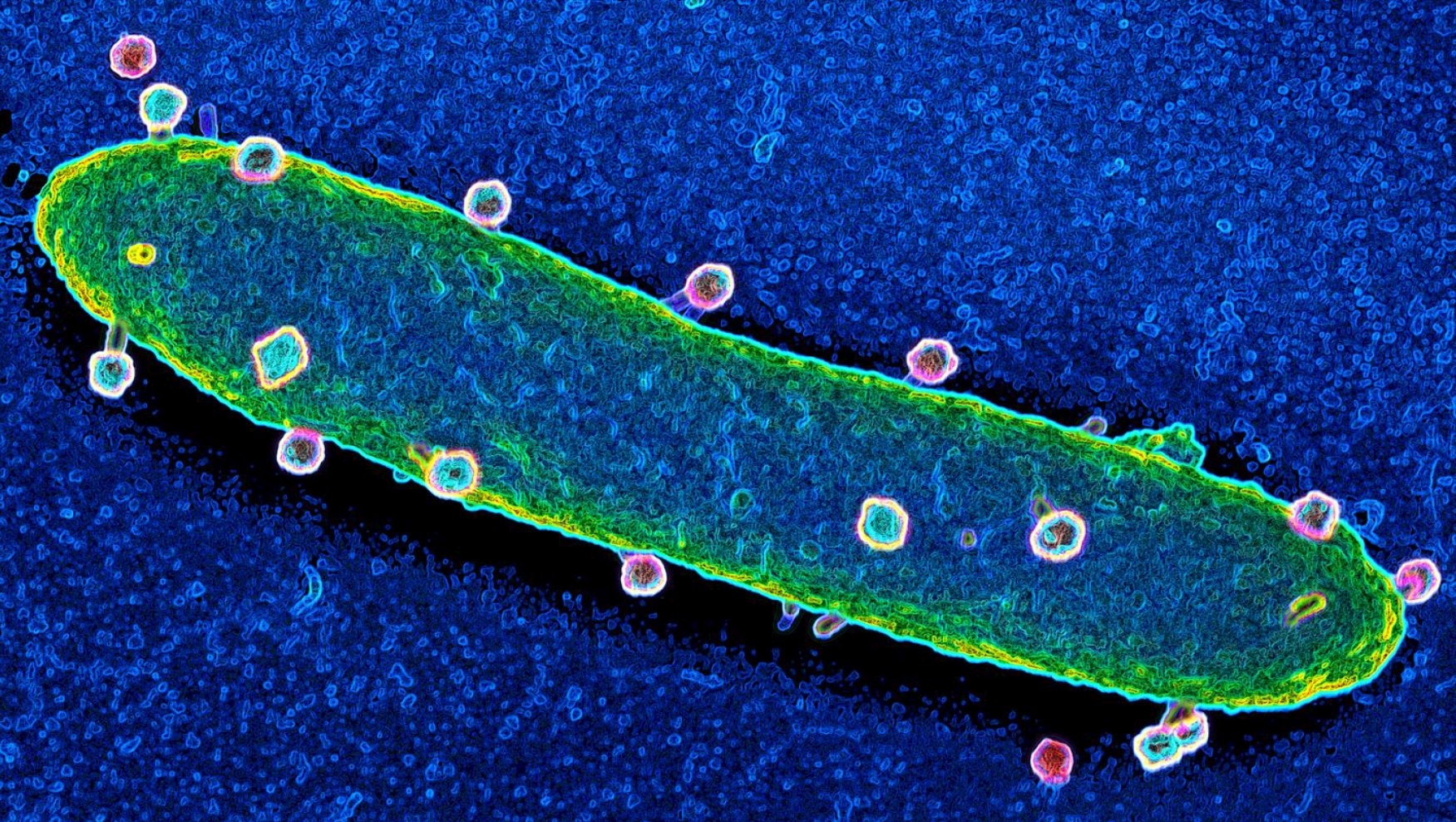
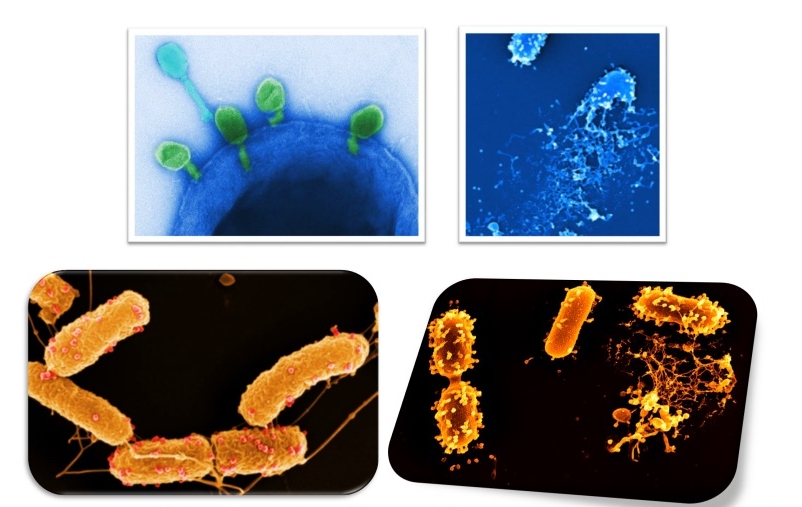
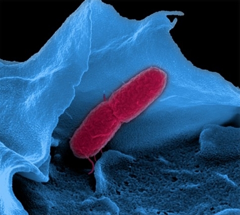
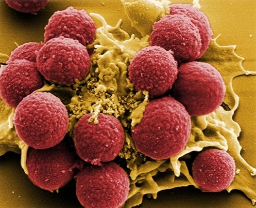
Both light- and electron-microscopy are ideally equipped to visualise the tiniest substructures of objects at the highest resolution. It can thus impressively visualise, for instance, how pathogenic bacteria attach themselves to or penetrate cells, as well as depict the virulence of bacteriophages. High resolution microscopy is the only possibility of investigating these processes morphologically and contributing to solving the mechanisms of pathogenicity.
By means of exhibits on display, visitors could see for themselves the working methods and preparations used in electron microscopy, giving an idea of the enormous effort required to visualise such small structures that are invisible to the eye.
If I understood correctly, all three scientists in the exhibition also approached the topic of cell vitality artistically. Were you, Heinrich Lünsdorf and Manfred Rohde already involved in art?
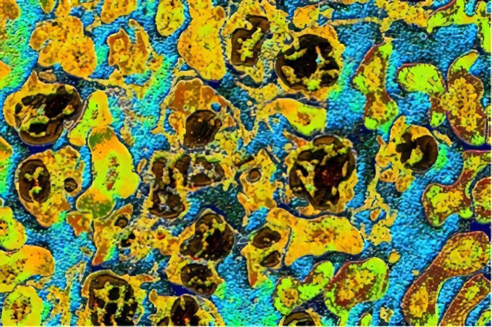
Lünsdorf and Rohde did not take an artistic approach in the stricter sense when alienating found structures, but they did use artificial colouring to highlight specific structures in the micrographs, which are necessary for the biological processes. I focused on biological forms and molecular signaling and tried to interpret them through painting.
Now to the artistic part of the exhibition: on the basis of your shared scientific knowledge on cells, did the three of you set yourselves certain artistic goals? Did you want to show something in particular through artistic means?
We were interested in pointing out mechanisms in a variety of forms and to shed light on energetic processes in the cell.
What was on display in the exhibition Cell Vitality?
In the entrance area, we exhibited particular aspects of the molecular dogma. The fundamental discoveries were presented so that the joy of discovery, creativity and the engagement of the scientists came to the fore. We also made clear that genetic engineering is a particular science with special experimental procedures.


Further into the exhibition, much space was devoted to the connections between the themes health/illness and their molecular processes. Original images of our own cell biological work was shown in combination with paintings.
We presented structures in the micro-range. Electron microscopic images of bacteria depicted forms that are so diverse and unusual, that they have no equivalent in the visible macroscopic world that surrounds us. We presented our own scientific results of the infection process caused by bacteriophages. Bacteriophages are bacteria-specific viruses that depend on bacteria in order to reproduce and eventually destroy the bacterium within a few hours. Virulent phages can be used as a new approach to combatting antibiotic-resistant bacteria. Impressive images show the bacterium’s attachment, multiplication and lysis (dissolution) process, as well as the release of multiple phages.
On the topic of tumour therapy, we presented two strategies that are based on changes in the immune cell. Our work in cell biology was exhibited in combination with intuitively painted objects. This presentation was regarded a radical challenge in the search for shared interfaces between science and art.


We chose cell receptors as the final exhibition theme. A receptor is a protein or protein complex that accepts specific signal molecules in a key-lock principle that induces specific processes in the cell. Our five senses rely on the function of specific receptors in order to receive the appropriate signals from our surroundings and to transmit these to the interior of the cell for further processing (light, sound, smell, taste, touch). Using the corona virus SARS-CoV-2 as an example, the binding of a surface protein (spike protein) to a cell receptor results in the penetration of the virus into the cell.


Earlier you indicated that you have also used artistic means (in a broader sense) to communicate findings of genetic engineering to a wider audience. What did you mean? How do you really relate artistic struggle with the topic cell vitality?
Lünsdorf and Rohde used colour to dye specific structures in their microscopic images in order to highlight those parts that are relevant and required for the biological function. The purpose of the dye is to draw the viewer’s attention.
I, on the other hand, concentrated on biological forms and signals at the molecular level and asked myself: What possibilities can I deploy to broaden the realm of visual art?
For the questioning of molecular objects and signals and finding an artistic response, a one-sided stance of figurative or abstract depictions was no longer considered relevant. Surreal, visionary and abstract ideas were approved. The image should have a life of its own that is not closely linked with the volitional formations or constraints of society. The viewer should get a feeling for the richness of nature as can be captured by genetic engineering.
So your objective was to point towards molecular mechanisms based on your scientific knowledge and through the use of artistic means in the broader sense and, as a result, to reflect on our relationship with nature, ourselves and our future. Thank you for this insightful interview.
Acknowledgements
I thank Dr. Johannes Urban Mayer, Uni-Marburg for advice on the topic of science communication, Dr. Victor Wray, HZI Braunschweig for discussion and Rebecca Grundmann for the revised translation.
References
Kunstforum International vol. 157 November – December 2001
Kunstforum International vol. 158 January – March 2002
https://wellcomecollection.org/articles/XcK2RBIAACMAXzcI
https://wellcomecollection.org/articles/W9b0kRIAABdu8KBo
https://niwa.co.nz/news/summer-series-2018-the-science-of-art-or-the-art-of-science https://cosmosmagazine.com/society/when-science-meets-art
https://edgy.app/where-art-and-science-intersect
https://theartofeducation.edu/2017/10/26/11-fascinating-artists-inspired-science/
Spektrum der Wissenschaft
Wissenschaft im Dialog
Bild der Wissenschaft
https://www.helmholtz.de/aktuell/presse-und-medien/mediathek/wissenschaftsbild-des-monats/
Details of the cover photo: Hubert Mayer: Skizze (2020). Photo: Hubert Mayer.
How to cite this article
Hubert Mayer and Peter Tepe (2022): Genetic Engineering and the Exhibition "Cell Vitality". w/k–Between Science & Art Journal. https://doi.org/10.55597/e8183

… [Trackback]
[…] Find More here to that Topic: between-science-and-art.com/genetic-engineering-and-the-exhibition-cell-vitality/ […]
… [Trackback]
[…] Find More Information here to that Topic: between-science-and-art.com/genetic-engineering-and-the-exhibition-cell-vitality/ […]
… [Trackback]
[…] Find More on to that Topic: between-science-and-art.com/genetic-engineering-and-the-exhibition-cell-vitality/ […]